Decoding Your 12-Week Ultrasound: A Comprehensive Guide
The 12-week ultrasound is a significant milestone in pregnancy, offering the first glimpse of your developing baby. Understanding what you’re seeing in those fuzzy images can be both exciting and reassuring. This comprehensive guide will delve into the details of a 12-week ultrasound, explaining what’s considered normal, what measurements are taken, and what to expect during the procedure. We aim to empower you with the knowledge to interpret your ultrasound results and engage in informed conversations with your healthcare provider. This isn’t just about pretty pictures; it’s about assessing your baby’s health and development.
Understanding the Purpose of a 12-Week Ultrasound
The 12-week ultrasound, also known as the nuchal translucency (NT) scan, serves several crucial purposes. While offering a precious first look at your baby, it’s primarily a screening tool designed to assess the risk of certain chromosomal abnormalities. This ultrasound typically takes place between 11 weeks and 13 weeks 6 days of gestation.
The primary goals of the 12-week ultrasound include:
- Confirming the pregnancy and viability: Ensuring that there is a heartbeat and that the pregnancy is progressing as expected.
- Determining gestational age: Accurately dating the pregnancy, which is essential for tracking development and predicting the due date.
- Assessing the number of babies: Identifying whether it’s a singleton, twin, or multiple pregnancy.
- Evaluating the nuchal translucency: Measuring the fluid-filled space at the back of the baby’s neck to assess the risk of Down syndrome (Trisomy 21), Trisomy 18 (Edwards syndrome), and Trisomy 13 (Patau syndrome).
- Early anatomical survey: Checking for the presence of major anatomical structures, such as the brain, spine, limbs, and abdominal wall.
It’s important to remember that the 12-week ultrasound is a screening test, not a diagnostic one. If the results indicate an increased risk of chromosomal abnormalities, further diagnostic testing, such as chorionic villus sampling (CVS) or amniocentesis, may be recommended.
What Does a Normal 12-Week Ultrasound Picture Show?
A normal 12-week ultrasound picture reveals several key features of your developing baby. While the images may appear somewhat indistinct to the untrained eye, a trained sonographer can identify various anatomical structures and assess their development.
Here’s what you can typically expect to see in a normal 12-week ultrasound picture:
- The baby’s overall shape and position: The baby will appear as a small, curled-up figure within the amniotic sac. The sonographer will assess the baby’s position to obtain accurate measurements.
- The head and brain: The head will be clearly visible, and the sonographer will check for the presence of the two brain hemispheres.
- The spine: The spine will appear as a line of small dots running down the baby’s back.
- The limbs: The arms and legs will be visible, and the sonographer will count the fingers and toes (although they may be difficult to see clearly at this stage).
- The abdominal wall: The sonographer will check that the abdominal wall is intact and that the internal organs are developing properly.
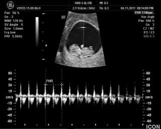
- The heart: The heart will be visible as a small, flickering structure. The sonographer will measure the heart rate to ensure that it’s within the normal range (typically between 120 and 180 beats per minute).
- The nuchal translucency: This is the fluid-filled space at the back of the baby’s neck. The sonographer will measure the thickness of this space to assess the risk of chromosomal abnormalities.
- Placenta and amniotic fluid: The ultrasound will also show the location of the placenta and the amount of amniotic fluid surrounding the baby.
It’s important to note that the appearance of the ultrasound picture can vary depending on the baby’s position, the quality of the ultrasound equipment, and the sonographer’s skill. If you have any concerns about the appearance of your ultrasound picture, don’t hesitate to discuss them with your healthcare provider.
Deciphering Key Measurements and What They Mean
The 12-week ultrasound involves several key measurements that provide valuable information about your baby’s development and health. Understanding these measurements can help you better interpret your ultrasound results and engage in informed discussions with your healthcare provider.
Crown-Rump Length (CRL)
The crown-rump length (CRL) is the measurement of the baby’s length from the top of the head (crown) to the bottom of the buttocks (rump). This is the most accurate measurement for determining gestational age in the first trimester. A normal CRL at 12 weeks is typically between 5.4 cm and 7.0 cm.
Nuchal Translucency (NT)
The nuchal translucency (NT) is the measurement of the fluid-filled space at the back of the baby’s neck. An increased NT measurement can indicate an increased risk of Down syndrome, Trisomy 18, or Trisomy 13. However, it’s important to remember that an increased NT measurement doesn’t necessarily mean that the baby has a chromosomal abnormality. The NT measurement is combined with the mother’s age and blood test results to calculate a risk assessment. A normal NT measurement at 12 weeks is typically less than 2.5 mm, but this can vary slightly depending on the laboratory.
Heart Rate
The baby’s heart rate is measured to ensure that it’s within the normal range. A normal heart rate at 12 weeks is typically between 120 and 180 beats per minute.
Other Measurements
In addition to the CRL, NT, and heart rate, the sonographer may also take other measurements, such as the biparietal diameter (BPD) of the head and the femur length (FL). These measurements can provide additional information about the baby’s growth and development.
Factors Influencing the Clarity of Ultrasound Pictures
Several factors can influence the clarity and quality of ultrasound pictures. Understanding these factors can help you manage your expectations and appreciate the limitations of ultrasound technology.
- Maternal body mass index (BMI): Women with a higher BMI may have less clear ultrasound images due to increased tissue density.
- Baby’s position: The baby’s position in the womb can affect the visibility of certain anatomical structures. If the baby is lying in an awkward position, the sonographer may have difficulty obtaining clear images.
- Amniotic fluid volume: The amount of amniotic fluid surrounding the baby can also affect image clarity. Adequate amniotic fluid helps to transmit the ultrasound waves and produce clearer images.
- Ultrasound equipment: The quality of the ultrasound equipment can significantly impact image resolution. Newer, more advanced equipment typically produces clearer images.
- Sonographer’s skill: The sonographer’s experience and skill in performing and interpreting ultrasounds are crucial for obtaining accurate and informative images.
What Happens if the Ultrasound Reveals Abnormalities?
If the 12-week ultrasound reveals any potential abnormalities, it’s natural to feel anxious and concerned. However, it’s important to remember that an abnormal ultrasound finding doesn’t necessarily mean that your baby has a problem. In many cases, further testing is needed to confirm or rule out any potential issues.
Common follow-up tests after an abnormal 12-week ultrasound include:
- Non-invasive prenatal testing (NIPT): NIPT is a blood test that screens for chromosomal abnormalities, such as Down syndrome, Trisomy 18, and Trisomy 13. NIPT is highly accurate and can be performed as early as 10 weeks of gestation.
- Chorionic villus sampling (CVS): CVS is a diagnostic test that involves taking a small sample of tissue from the placenta. CVS can be performed between 10 and 13 weeks of gestation and can detect chromosomal abnormalities and certain genetic disorders.
- Amniocentesis: Amniocentesis is a diagnostic test that involves taking a small sample of amniotic fluid. Amniocentesis is typically performed between 15 and 20 weeks of gestation and can detect chromosomal abnormalities, genetic disorders, and neural tube defects.
- Detailed ultrasound: A more detailed ultrasound, typically performed around 20 weeks of gestation, can provide a more comprehensive assessment of the baby’s anatomy and development.
Your healthcare provider will discuss the specific findings of your ultrasound and recommend the most appropriate follow-up tests based on your individual circumstances. It’s important to ask questions and express any concerns you may have. Remember that you are not alone, and your healthcare team is there to support you throughout your pregnancy.
The Emotional Impact of Seeing Your Baby on Ultrasound
The 12-week ultrasound is often a deeply emotional experience for expectant parents. Seeing your baby for the first time can evoke a range of feelings, from joy and excitement to awe and wonder. The ultrasound picture provides a tangible connection to your developing baby and can strengthen the bond between parents and child.
For some parents, the ultrasound can also be a source of anxiety and stress, especially if there are any concerns about the baby’s health. It’s important to acknowledge and validate these feelings and to seek support from your partner, family, friends, or healthcare provider.
Remember that the 12-week ultrasound is just one step in the journey of pregnancy. There will be many more milestones and experiences to come. Take the time to cherish this special moment and to celebrate the miracle of life.
Expert Perspectives on Ultrasound Technology Advancements
Ultrasound technology is continuously evolving, leading to more detailed and accurate imaging. Experts in the field emphasize the importance of staying current with these advancements to provide the best possible care for pregnant women and their babies. New technologies, such as 3D and 4D ultrasounds, offer even more realistic views of the developing baby and can aid in the early detection of certain abnormalities.
These advancements not only improve diagnostic capabilities but also enhance the emotional experience for parents, allowing them to connect with their baby in a more profound way. As technology continues to advance, we can expect even more sophisticated ultrasound techniques to emerge, further improving prenatal care and outcomes.
Making the Most of Your 12-Week Ultrasound Experience
The 12-week ultrasound is a significant event in your pregnancy journey, offering valuable insights into your baby’s development and health. By understanding what to expect during the procedure, what measurements are taken, and what the results mean, you can approach your ultrasound with confidence and knowledge. Remember to ask questions, express any concerns, and cherish this special moment as you connect with your developing baby. With careful preparation and a clear understanding of the process, you can transform your 12-week ultrasound into a positive and empowering experience.
